Research

The endolysosomal/Golgi system provides routes for degradation or recycling of plasma membrane (PM) proteins via endocytosis and degradation of cytosolic proteins/organelles via autophagy. Much evidence supports the idea that endolysosomal system dysfunction underlies a variety of neurodegenerative conditions, ranging from Alzheimer’s Disease to Parkinson’s diseases. Among the proteins encoded by genetic drivers of these are related diseases are core components of the endolysosomal system itself (e.g. BIN1, PSEN1/2, GRN, CHMP2B, VPS35, LRRK2) and proteins whose endolysosomal trafficking is disrupted in the context of genetic disease (APP/Abeta, SORL1, Tau, alpha-synuclein aggregates). Nevertheless, how individual risk alleles alter trafficking and degradation pathways within the network and whether diverse alleles converge on a common set of phenotypic outcomes remains unclear. This reflects, in part, the absence of a framework for understanding the key protein assemblies within the endolysosomal system that are subject to altered regulation in the context of risk alleles. We are using organelle isolation by immunoprecipitation (Endo-IP, Lyso-IP, Mito-IP) in the context of ES cell-derived iNeurons to systematically elucidate the proteome organization of organelles in various cell states and how protein interactions within these organelles are affected in the context of neurodegenerative disease or lysosomal storage disorder alleles. We have developed systems for systematic analysis of organelle proteomes using mass spectrometry (PNAS, 2024), and have created libraries of engineered cell lines including ES cells to facilitate an understanding of how organelle proteomes and interactions are altered in the context of specific mutations (BioRxiv, 2024; Science Advances, 2025). In this context, we are using single particle cryo-EM as well as in situ cryo-ET (including collaborations with the Wilfling and Schulman labs) to understand the structures of specific complexes and to understand organellar defects with nanometer resolution within cells.

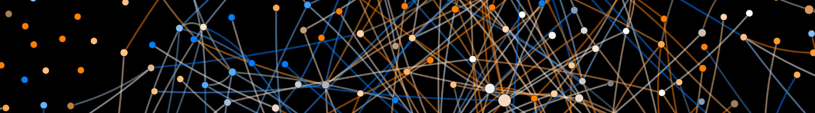
The human interactome is composed on thousands of interactions and protein communities. Using a robust interaction proteomics platform, we - together with the Gygi lab - have performed interaction proteomics on more than 17,000 human proteins in HEK293T cells and 10,000 proteins in HCT116 cells. This has led to now three iterations of the BioPlex database of interacting proteins (available at https://bioplex.hms.harvard.edu/) [Cell, 2015, Nature, 2017, Cell, 2021]. Current efforts seek to convert these interaction networks into structural models for the proteome using Alphafold, with more than 100,000 predictions performed thus far. The creation of a structural interactome will fuel numerous hypothesis-driven experiments to elucidate functions of poorly understood proteins, often referred to as the “dark proteome”.

CRLs represent the largest collection of E3 ubiquitin ligases and contain a cullin scaffold and one of more than 200 substrate specific adaptors. Our lab co-discovered these E3s together with the Elledge and Deshaies lab (Cell, 1996) and we have identified numerous targets and regulatory mechanisms over the years. We are using a variety of approaches to understand substrates and regulation of CRLs, including the development of “degradomic” approaches, in the context of changes in cell states. In addition, we are collaborating with the Schulman lab to use proteomics and structural biology for elucidation of molecular glues that function with distinct types of CRLs. We are exploring targets and regulation of FBXO7, and enigmatic CRL substrate adaptor that is mutated in familial forms of Parkinson’s disease, particularly its role in mitochondrial quality control (EMBO Reports, 2023).

A major effort in the lab relates to the development of methods for proteomic analysis of protein complexes and organelles, as well as regulatory modifications. Beyond BioPlex, we are developing quantitative proteomics approaches to elucidate targets of E3 ubiquitin ligases using Kgg proteomics, as we have done extensively for Parkin in the context of mitophagy (Nature, 2013; Mol Cell, 2018, 2019). In addition, we are developing global proteomics workflows to understand large-scale proteome alterations in response to specific signals (nutrient stress, changes in developmental state, etc) (Nature, 2023; Mol Cell, 2021). More recently, we have developed approaches for isolation of organelles such as early endosomes (Nature Communications, 2021; PNAS, 2024), allowing us to identify resident endosomal proteins as well as cargo for endocytosis. We are using these approaches, coupled with cross-linking mass spectrometry and correlation profiling based approaches to identify molecular assemblies on intact organelles, ranging from endosomes to Golgi. We are combining this with Alphafold to predict structures for poorly understood complexes as a starting point for functional studies. This is providing what we refer to as structural proteome landscapes for individual organelles, which we are further interrogating via biochemical and structural approaches.
We are grateful for the generous support of our funding agencies!